VisArthro: Computer Vision For Computer Aided Arthroscopy
Project name: VisArthro: Computer Vision For Computer Aided Arthroscopy
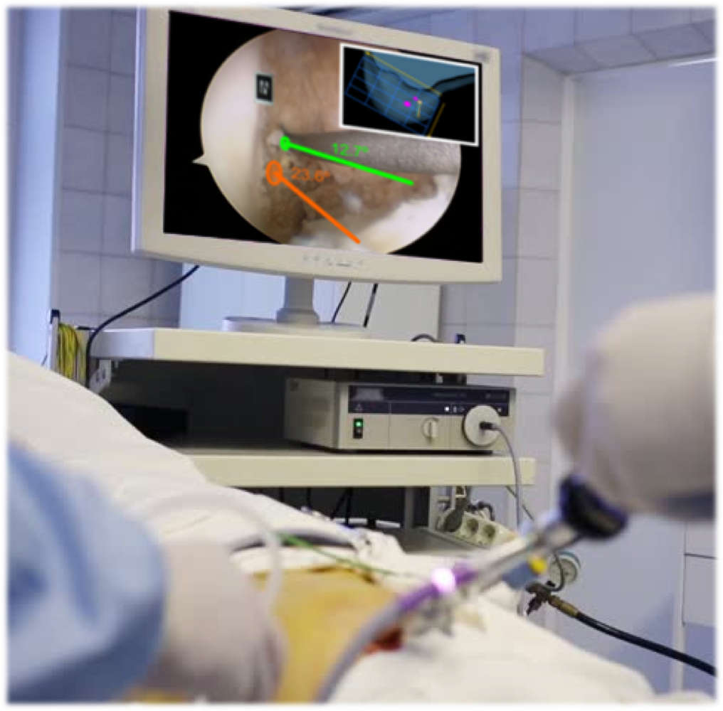
Description: Arthroscopy is a Minimally Invasive Surgical (MIS) procedure for treatment of damaged joints in which instruments and endoscopic camera (the arthroscope) are inserted into the articular cavity through small incisions (the surgical ports). Arthroscopic procedures are very difficult to execute because of indirect visualization and limited manoeuvrability inside the joint, with novices having to undergo a long training period and experts often making mistakes of clinical consequences. The project aims to overcome this drawback by conducting research in computer vision techniques to build a system for Computer-Aided Arthroscopy (CAA) that will guide the surgeon throughout the procedure. Such system will exclusively rely in processing the arthroscopic video, being the first of the kind not requiring additional intra-operative sensing modalities such as opto/magnetic tracking. This is a crucial feature to assure broad adoption among surgeons, ultimately contributing to disseminate and improve the clinical outcome of arthroscopy, with indisputable benefits for the patient. Our research will be motivated by two specific procedures: the reconstruction of the Anterior Cruciate Ligament (ACL) in the knee and the removal of Femur Acetabular Impingements (FAI) in the hip. ACL tear is a common pathology for which arthroscopy is the standard treatment (> 300000 cases per year worldwide). The procedure consists in replacing the torn ACL by a substitution graft that is pulled into the joint through a tunnel opened with a drill (Fig. 1). Placing this tunnel in the correct anatomical position is crucial for the knee to fully recover its functionality [1,2]. Recent studies show that the dominant surgical technique, the transtibial approach, only reaches optimal tunnel placement in 61% of the cases [1]. FAI occurs when the ball shaped femoral head rubs abnormally in the acetabular socket, which in 91% of the cases is caused by an excess of bone tissue in the femur head-neck that creates a bump known as CAM impingement [3] (see Fig 2). The treatment is surgical and consists in removing the CAM to restore the ball shape to the femur-head. Unfortunately, such reshaping via arthroscopy is extremely difficult to accomplish being only performed by a small number of highly skilled surgeons (e.g. in Portugal there are only two surgeons doing the procedure).
PI: João Pedro Barreto
Reference: PTDC/EEI-AUT/3024/2014
Website: home.deec.uc.pt/~jpbar/visarthro/
Fundind entity: FCT
Budget: 200K€
Duration of the action: 2016-07-01 - 2019-06-30